
LIPOMAS AND THEIR SURGICAL TREATMENT
Lipomas are benign tumors of the fatty tissue. They are generally well-defined because they are surrounded by a thin tumor capsule. Usually, they can be easily felt and moved against the surrounding tissue. Lipomas are almost always soft and painless. However, if they are angiolipomas (lipomas with an increased blood vessel component), they can be typically more painful.
OP-PHOTOS
Would you like to see them?
Why do we show you surgical photos?
Many people believe that surgeries are robust, blood-rich, and chaotic.
Not with us!
We operate delicately, gently, with very little blood, and with the best possible overview,
displaying all anatomical structures optimally.
We want to showcase this.
This is our surgical quality.
And this is also the reason why we have an almost "zero" complication rate in surgeries.
We aim to provide the best possible transparency for visitors to our website.
Decide for yourself, whether you would like to see these surgical photos / films.
Go to the surgical photos!
Lipomas can occur in various parts of the body. They are commonly found wherever there is fatty tissue, such as on the trunk and limbs beneath the skin. However, in rarer cases, lipomas have been found in extraordinary locations in the body, including the spinal cord and the brain.
They can occur as single tumors or in the form of numerous tumors scattered throughout the entire body, sometimes numbering in dozens or even hundreds. Often, there is a family history of lipomas, meaning that one of the parents also suffers from lipoma tumors.
Lipomas, being benign tumors, are not inherently dangerous. Therefore, many doctors advise against surgery, especially when they are small. At the beginning, they tend to grow slowly. However, as they get larger, their growth accelerates because the cells have the potential to divide: If 8 cells divide gradually, they become 16 cells. But if 10,000 cells divide in the same period, they become 20,000 cells. This means that the overall growth of the lipoma tumor increases rapidly. Extreme cases from India are known where lipomas weighed up to 20 kg, for example, on the back. In my own patient cases, I have encountered lipomas the size of a handball, which is already burdensome enough.
Repressive Lipoma Growth
Lipoma Growth Pressure
Lipomas have a dangerous characteristic that should be taken seriously: they exert growth pressure. The dividing cells naturally require more space and continue to divide further. Like an expanding balloon, the tumor grows in size. This expansion occurs more easily in areas that exert less counter-pressure. Generally, these are blood vessels and nerve pathways that utilize natural openings (weak points) in the body tissues for their passage. As a result, there are repeated instances of tapering growth of lipoma portions into the blood vessels and nerve pathways, where they cause displacements only in the later stages. Because of this property, lipomas are often underestimated. To be radically removed, nerves and blood vessels often need to be extensively exposed microsurgically, not without the risk of injuring these important structures. This requires a great deal of surgical skill and experience to completely remove such tumors.
Therefore, after many years of surgical practice, one thing I have undoubtedly learned is that lipomas are almost always underestimated, even by experienced surgeons. This includes myself, despite all my experience.
Another peculiarity of lipomas is that the larger they become, the more they divide into lobe-like substructures. They do not remain round but rather become fragmented. This makes their surgical removal quite tricky.
Example of a 68-year-old female patient with a large lipoma of the right elbow
Photos: Dr. Roman Fenkl, with the written consent of the patient
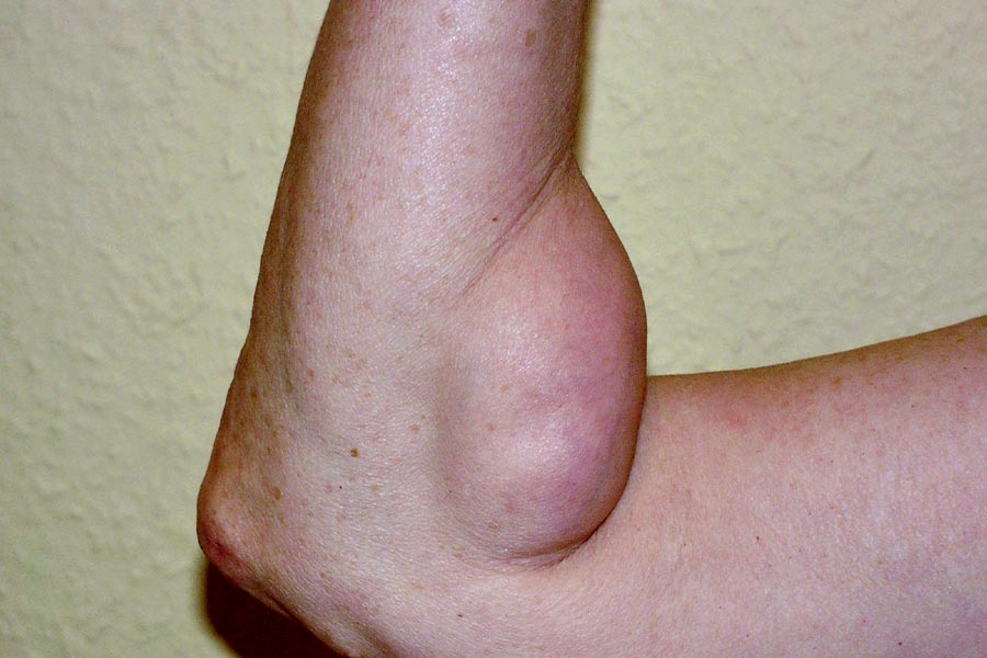
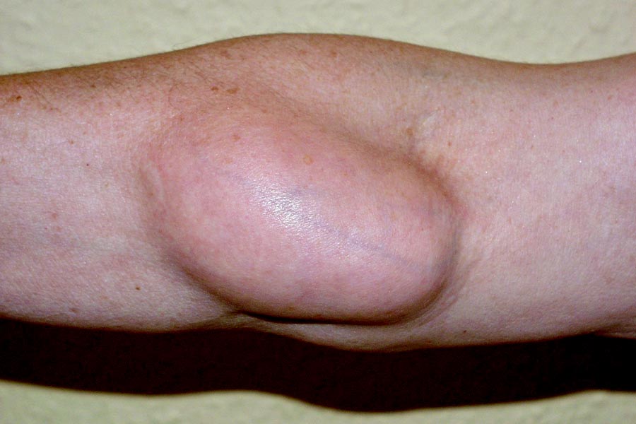
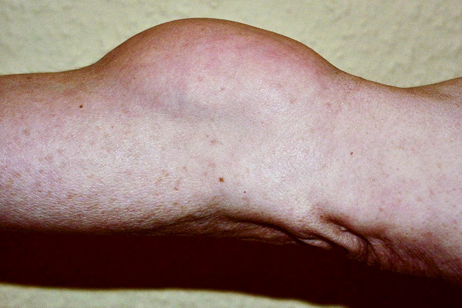
Large lipoma in the right elbow of a 68-year-old patient. The general practitioner had advised against surgery for years, as the growth was deemed "non-threatening." However, this changed when it began causing mechanical issues with elbow flexion and difficulties wearing long-sleeved clothing. Additionally, tingling sensations started occurring in the hand, accompanied by partial numbness in the fingers. The growth continued to expand repressively into the main compartment of major blood vessels and nerves in the elbow.
OP-PHOTOS
Would you like to see them?
Why do we show you surgical photos?
Many people believe that surgeries are robust, blood-rich, and chaotic.
Not with us!
We operate delicately, gently, with very little blood, and with the best possible overview,
displaying all anatomical structures optimally.
We want to showcase this.
This is our surgical quality.
And this is also the reason why we have an almost "zero" complication rate in surgeries.
We aim to provide the best possible transparency for visitors to our website.
Decide for yourself, whether you would like to see these surgical photos / films.
Go to the surgical photos!
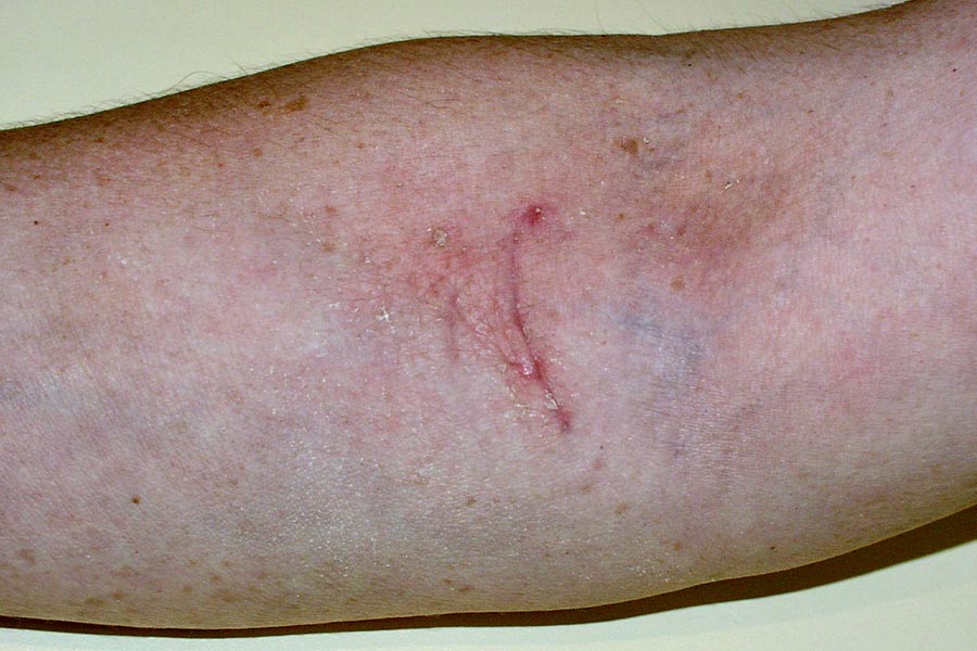
The healing outcome with scar 8 weeks postoperatively.
The scar is still red and will gradually fade. It can be observed that the scar is significantly smaller than the lipoma itself (compare with preoperative findings).
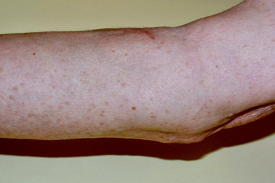
When viewed from the side, the skin surface of the forearm and elbow is even, not indented as in its natural state. The body's integrity has been restored. This could only be achieved through the intraoperative filling of the tissue displacement defect using two fat flap grafts, one from above (from proximal to distal), and the other from below (from distal to proximal).
Satellite Lipomas
Furthermore, lipomas tend to form so-called satellite lipomas. These are often tiny, smaller lipomas that develop in the immediate vicinity of the main lipoma, likely branching off from the primary tumor and becoming independent. If the surgical procedure is not performed with extreme precision and attention, these small satellite growths can be easily overlooked as they may resemble natural fatty tissue. If not removed during the operation, it is likely that they will develop into a new lipoma tumor over time, known as a lipoma recurrence. Recurrences are often accepted as "inevitable," but in reality, the main reason is almost always a lack of thorough preparation (excision) of the primary tumor. This is not surprising, as health insurance companies generally do not prefer to cover the costs of a surgeon taking the time to carefully and meticulously dissect along the tumor capsule. Surgeons tend to favor the "5-minute technique," where the lipoma is swiftly encircled with a finger during the operation and "ripped out" in this manner. Needless to say, in such cases, recurrences are simply a matter of course. However, private health insurance companies also don't like to pay for more, as they classify lipomas as "trivial, harmless, benign tumors of fatty tissue." Unfortunately.
OP-PHOTOS
Would you like to see them?
Why do we show you surgical photos?
Many people believe that surgeries are robust, blood-rich, and chaotic.
Not with us!
We operate delicately, gently, with very little blood, and with the best possible overview,
displaying all anatomical structures optimally.
We want to showcase this.
This is our surgical quality.
And this is also the reason why we have an almost "zero" complication rate in surgeries.
We aim to provide the best possible transparency for visitors to our website.
Decide for yourself, whether you would like to see these surgical photos / films.
Go to the surgical photos!
Skin indentations postoperatively, after lipoma removal
I frequently encounter patients who had larger and smaller lipomas removed elsewhere and now have deep dents at the sites of lipoma excision. Such "holes" can be aesthetically very unpleasant, but they are also a consequence of "budget medicine." A lipoma that was allowed to gradually grow over the years occupies a certain space. When this lipoma is removed, the displaced healthy tissue does not simply snap back; it finds a new place. To avoid a crater-like depression under the skin closure, the surrounding tissue, mostly fatty tissue, must be brought back into the defect. This is surgically accomplished by mobilizing smaller or larger flaps of fatty tissue, which remain vascularized but are partially detached and shifted into the defect. They must be carefully sutured together to keep them in place. This is because the patient's movements during the first postoperative weeks can cause the "butter-soft" fatty tissue flaps to tear at their stitches and retract, thereby revealing the original "dent" again. Therefore, a postoperative period of rest is necessary. Such fatty tissue flap procedures are not simple surgeries and require a certain level of plastic surgical experience. The difficulty of this surgical technique lies in the fact that if the operation is successful, nothing should be visible; the skin surface should be even, which is what one would intuitively expect after surgery. I usually discuss with my lipoma patients the necessity of such fatty tissue flap procedures for a better aesthetic outcome. If the decision is against it, usually due to cost reasons, the operation is also simpler and much shorter. Here, the necessity of surgical intervention lies in the eye of the beholder, in the avoidance of disfigurement.
Example of a 52-year-old female patient after the removal of a large lipoma on the outside of the hip
Photos: Dr. Roman Fenkl, with the written consent of the patient
A 52-year-old patient, years after the surgical removal of a larger lipoma on the outer side of the hip ("saddlebags") performed by a general surgeon elsewhere. It's clear to see that the doctor took great care with the aesthetic stitching (so-called intracutaneous sutures).
However, a noticeable indentation of the body surface is quite evident, resulting from the fact that the surgeon did NOT fill the excision defect of the large lipoma intraoperatively through tissue displacement. I reevaluated this patient after 14.5 years and had to note that there has been no change or improvement in this indentation.
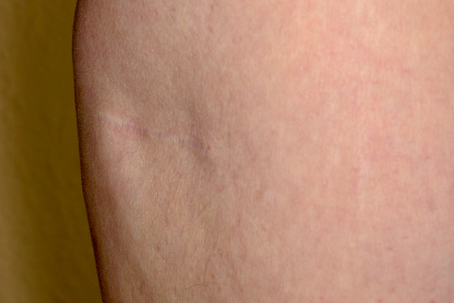
Previous findings (2006) viewed diagonally from the front.
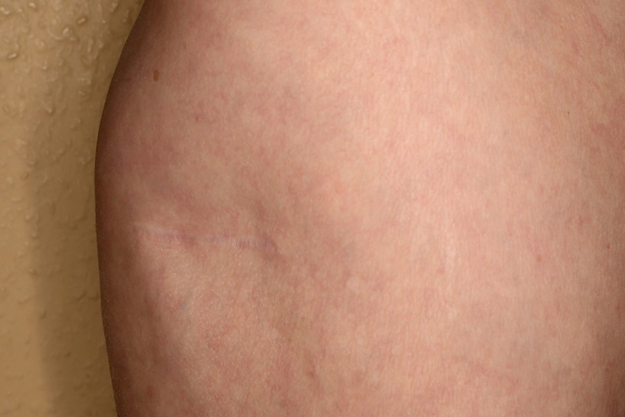
Current control findings 14.5 years later (2020). There has been no change in the indentation.
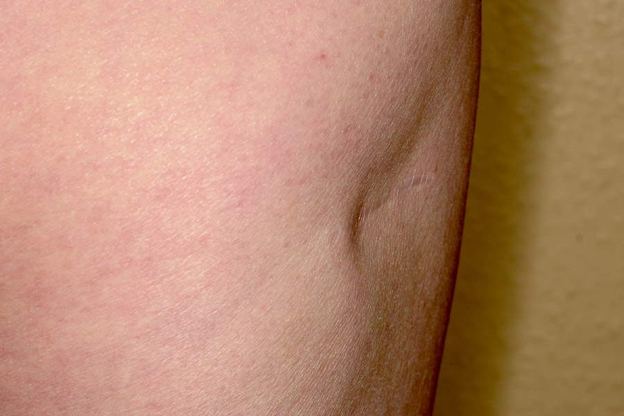
Previous findings (2006) viewed diagonally from the back.
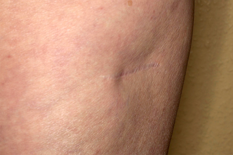
Current control findings 14.5 years later (2020). There has been no change in the indentation.
LIPOMA REMOVAL WITH INTRAOPERATIVE DENT FORMATION
Example of a 64-year-old female patient with a large lipoma on the left thigh
Photos: Dr. Roman Fenkl, with the written consent of the patient
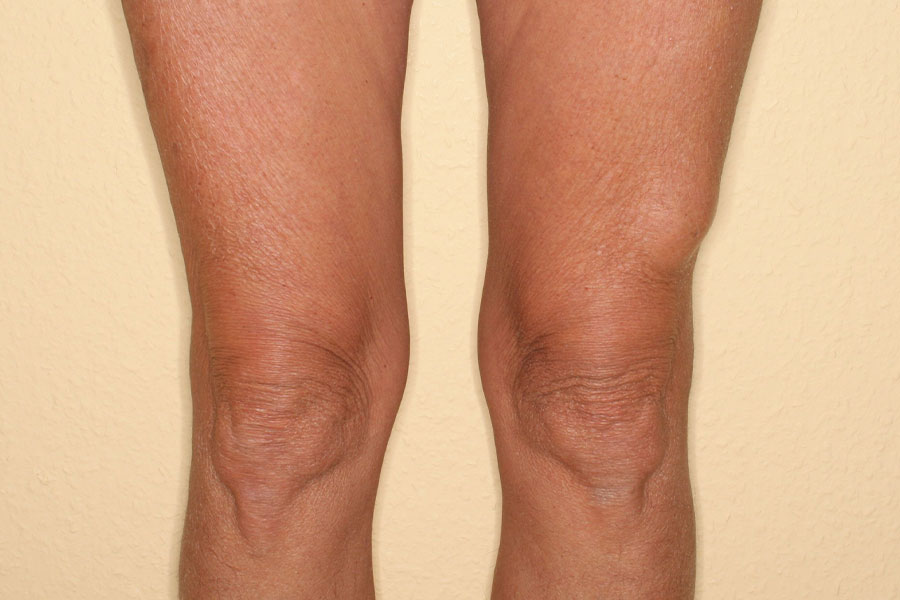
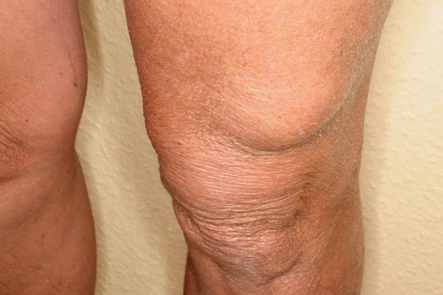
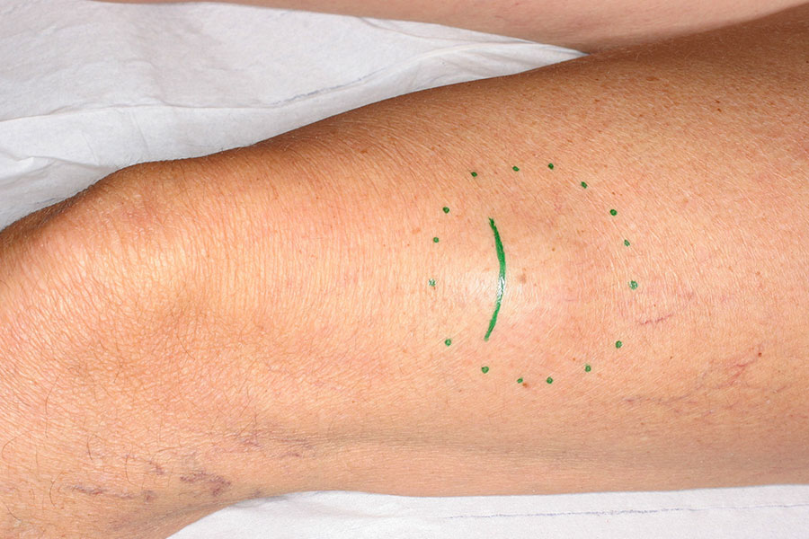
Photos 1 + 2: 64-year-old patient with a large lipoma on the distal front side of her left thigh, above the knee joint. Additionally, there is pronounced actinic skin damage (caused by excessive sun exposure) with loss of tension and elasticity of the skin.
Photo 3: Planning immediately before the operation. Dotted line: The presumed (palpable) boundaries of the growth. Solid line: The intended/planned incision line, smaller than the growth and with a curved path, following the discernible lines of skin creases (skin fold patterns, skin tension lines) for the purpose of optimizing the future scar.
OP-PHOTOS
Would you like to see them?
Why do we show you surgical photos?
Many people believe that surgeries are robust, blood-rich, and chaotic.
Not with us!
We operate delicately, gently, with very little blood, and with the best possible overview,
displaying all anatomical structures optimally.
We want to showcase this.
This is our surgical quality.
And this is also the reason why we have an almost "zero" complication rate in surgeries.
We aim to provide the best possible transparency for visitors to our website.
Decide for yourself, whether you would like to see these surgical photos / films.
Go to the surgical photos!
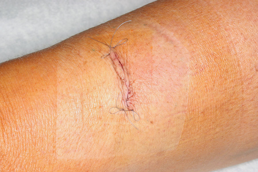
3 days postoperative,
aesthetic intracutaneous suture.
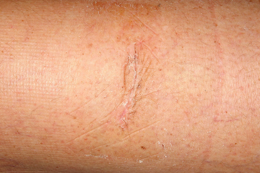
6 weeks postoperative
Due to the sun-damaged, tension-reduced skin, the scar healed particularly fine and inconspicuous. The patient did not seek further medical attention thereafter.
Special features of lipoma surgery
There is the rarer occurrence of lipomas that appear in the head region, preferably on the forehead or the hairy scalp. There, they cause bulging, aesthetically disturbing protrusions. If they are situated near nerve pathways, they can even trigger migraine-like headaches. Often, in such locations, lipomas are somewhat half-heartedly operated on because they sit very deep, mostly directly on the skull bone. Finding them there is much more challenging than one might assume, based on the seemingly "simple" external appearance. Time and again, patients who have been previously operated on visit my practice, with only part of the lipoma removed and complaints of remaining lipoma at the same site. The subsequent operation is possible, but due to the scar tissue that formed after the first, relatively easier operation, it is now significantly more difficult, especially if one wishes to keep the incision, for example, on the forehead, to a minimum..
Example Forehead Lipoma
In some individuals, there are numerous lipomas distributed over the body, sometimes hundreds. Removing each individual lipoma with separate skin incisions would mean disfiguring the entire body. Here, strategic operative planning and tactics come into play: Many lipomas group together in certain areas of the body. In such cases, strategically placed incisions can reach multiple lipomas through a single cut. It is possible, for example, to remove 4-5 lipomas through a single skin incision. This way, sometimes 30-50 lipomas can be removed in one session, on an outpatient basis under local anesthesia, but it requires considerable time.
Examples of multiple lipomas on the thigh
The most important questions before lipoma surgery
| How many lipomas can be removed in one session? |
The removal of (almost) all sizes of lipomas is usually done on an outpatient basis under local anesthesia, with additional sedation if necessary. Two very important factors play a crucial role here: |
| Can lipomas actually be suctioned (liposuctioned)? |
Lipomas consist of slightly (benign) degenerated fatty tissue. So, shouldn't they be able to be "easily and with minimal scarring" removed? |
| What are the risks of lipoma removal? |
Lipoma removals are generally safe and very low-risk procedures. One reason for this is that, from my experience: |
| Will the costs of lipoma removal be covered by health insurance? |
Public Health Insurance: Unfortunately, within our current German healthcare system, it does not matter whether one or 30 lipomas are removed in a single operation; the reimbursement remains the same. Therefore, such extensive cases can only be performed either through a special agreement with the public health insurance (GKV) or through separate private compensation. |
Overview of Surgical Therapies for Lipomas by Body Regions
Here you will find an overview by body regions of all the issues and treatment methods related to "Lipomas" described on this website.
Your Appointment in Our Practice
I would be pleased to meet you in my practice for a consultation in Darmstadt-Griesheim. Please book an appointment telephonically. Please mail us for further information under info(at)dr.fenkl.de.
For a personal consultation call the number below at any time between 08:00 – 18:00 from Monday to Thursday
Tel. 0049 6155 - 87 88 84

