NECK LIPOMAS
The neck is very complexly structured. There are many structures here that could be injured, including nerves and blood vessels, as well as many very delicate muscles and muscle attachments, lymph vessels and nodes, and organs such as the thyroid and parathyroid glands. Moreover, the "entrance" to the chest (mediastinum) is perilously close and easily accessible from here. Therefore, it often pays off to perform precise tumor localization with MRI (magnetic resonance imaging) before neck lipoma surgery. Furthermore, the very sensitive "neck" region cannot tolerate any contracting scar tissue that could disrupt future head posture and movement. As in the face, extremely careful surgery is a basic requirement here.
Example of a 55-year-old patient with a very large neck lipoma
Photos: Dr. Roman Fenkl, with the written consent of the patient
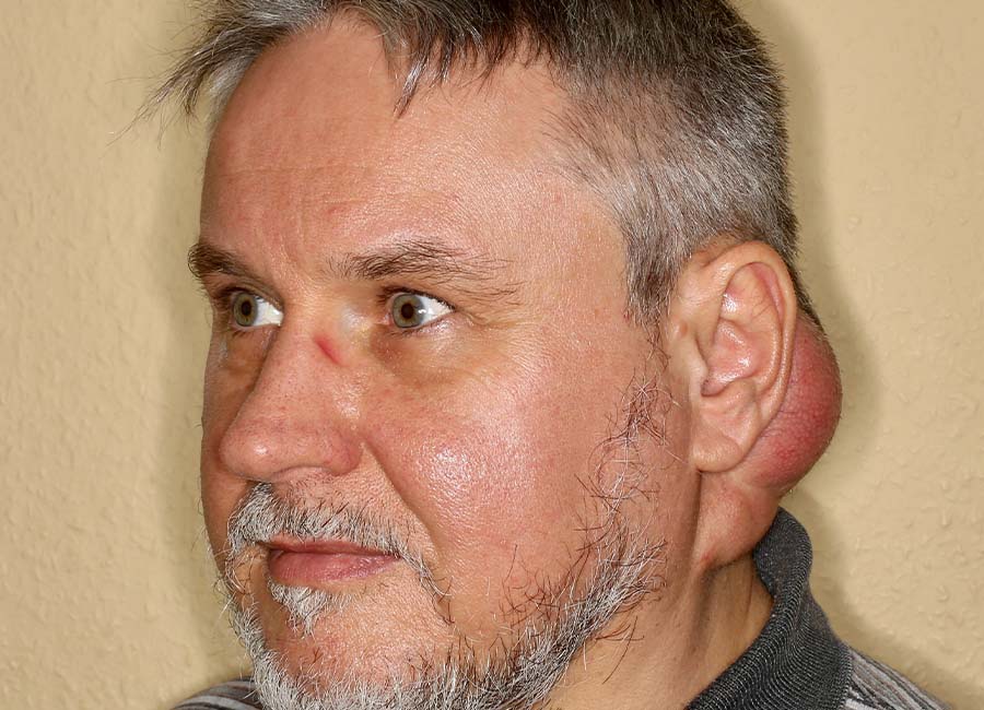
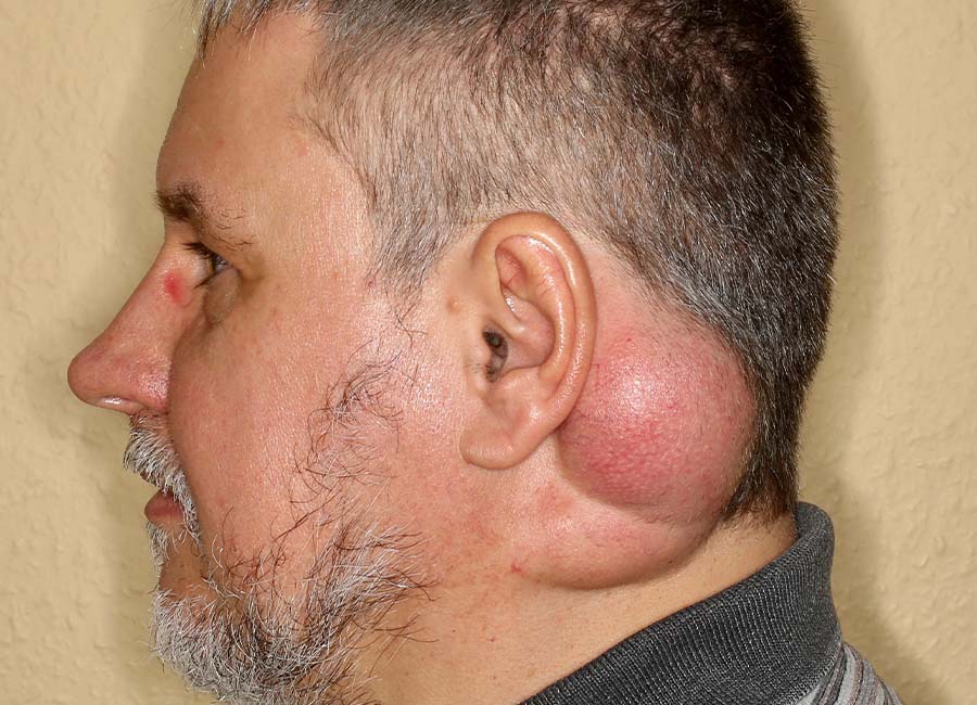
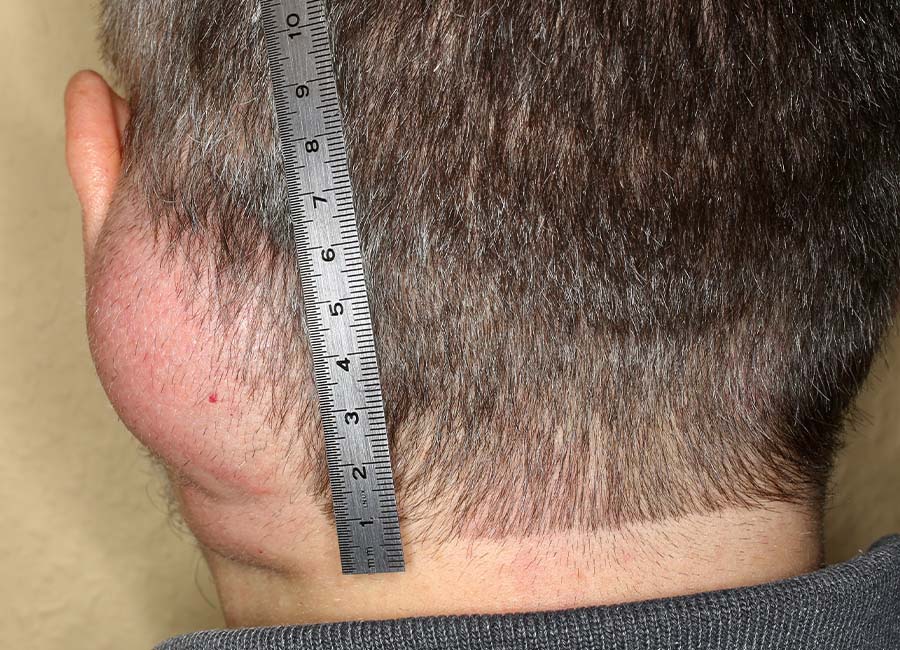
Photos 1 to 3: The initial condition. This 55-year-old, very amiable patient consulted us with a massive soft tissue mass behind his left ear that extended down to his neck. He reported that he had already entered first class of school with this – originally much smaller – mass, and had attended school, completed his entire education at university, and even married with the tumor. He had not experienced any neurological deficits, but his concerned wife, a nurse, had been encouraging him to have it removed. Eventually, he found our practice on the internet. The mass was clearly a benign, soft, but "gigantic" tumor, unmistakably a lipoma.
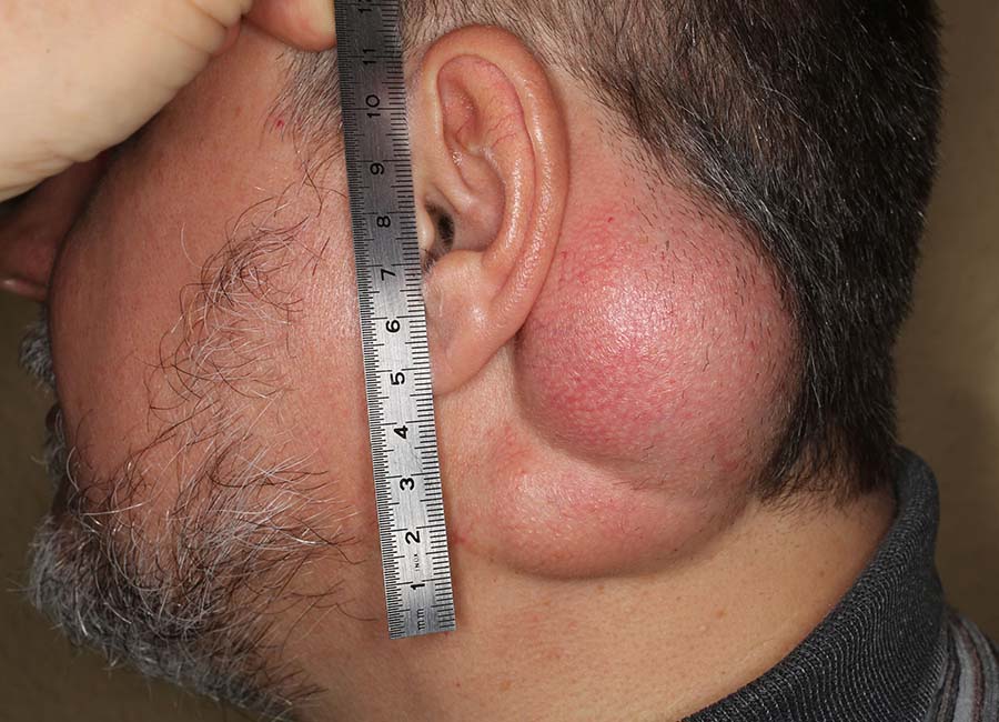
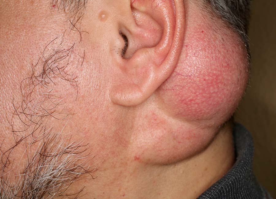
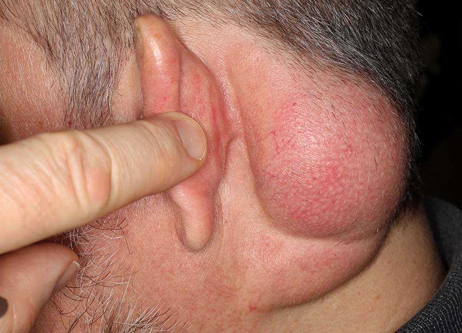
Photos 1 to 3: The initial condition. Upon closer examination, the lipoma occupied its place behind the ear and at the back of the head, extending its tendrils down to the neck, where it sat precisely on the most important ear nerve, the Nervus auricularis magnus. Here, the nerve surfaces from deeper tissues and is vulnerable to injury during surgical procedures, which is well-known to a plastic surgeon. Additional natural tissue structures confine this lipoma, making it appear divided into two parts. Behind the ear, the tumor extends right up to the earlobe – and thus, the delicate nerve and blood vessel structures of the earlobe.
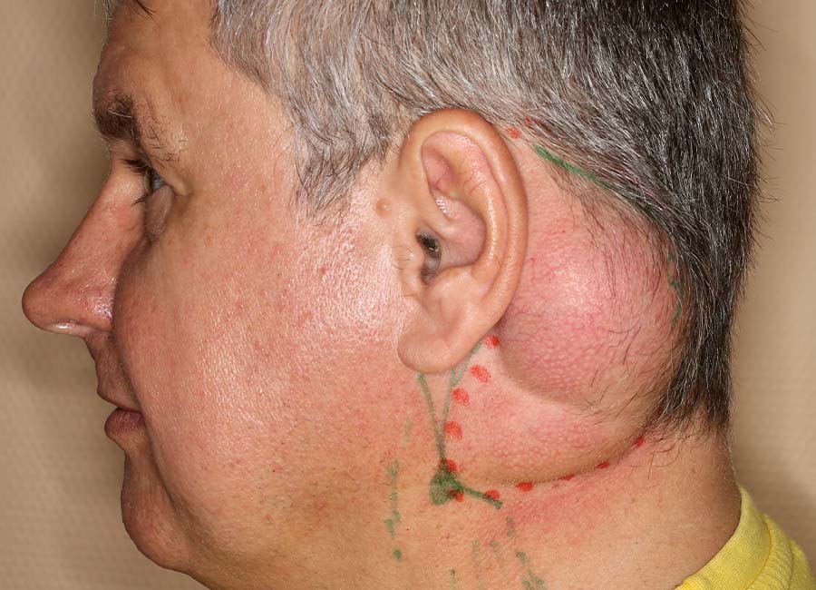
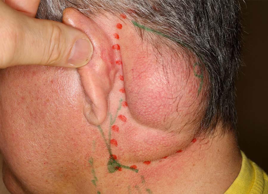
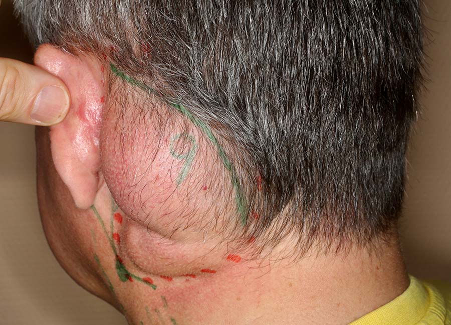
Photos 1 to 3: Preoperative surgical planning on the day of the operation. For this, the patient was asked to shave his full beard. The reason for this is the intention to maintain the utmost sterility during the surgical procedure. A full beard always carries additional microbes and thus serves as a potential source of infection. This is unnecessary for a procedure that is expected to be extensive. The tumor borders (red dots) are marked, along with the oblique neck muscle (M. sternocleidomastoideus) and the anatomically assumed exit point of the main ear nerve (N. auricularis magnus) with its branching nerves towards the ear. Intraoperatively, we could then "say hello" to this nerve. The surgical incision was deliberately made somewhat concealed at the hairline, taking into account aesthetic considerations.
OP-PHOTOS
Would you like to see them?
Why do we show you surgical photos?
Many people believe that surgeries are robust, blood-rich, and chaotic.
Not with us!
We operate delicately, gently, with very little blood, and with the best possible overview,
displaying all anatomical structures optimally.
We want to showcase this.
This is our surgical quality.
And this is also the reason why we have an almost "zero" complication rate in surgeries.
We aim to provide the best possible transparency for visitors to our website.
Decide for yourself, whether you would like to see these surgical photos / films.
Go to the surgical photos!


The postoperative course
after a 7-hour and 21-minute microsurgical operation under local anesthesia (the patient did not want or need any sedation, though it was offered by us).
Photo 1: The 1st day postoperative with the drainage (in yellow) still in place, which was painlessly removed. The expected postoperative swelling is still evident. The patient had no postoperative pain.
Photo 2: On the 6th day postoperative, the swelling has noticeably reduced but is still present. No pain is reported.


Photos centre:
On the 13th day postoperative, there is barely any sign of swelling. The patient was completely symptom-free but was advised to take it easy in terms of physical activity. Returning to work was allowed, but engaging in sports activities was still postponed.


Photos right:
15 months postoperative, there were no signs left of the lengthy operation. The patient was completely symptom-free, with no nerve deficits and no more swelling. For the remaining surgical scars, please refer to the following photos. The full beard has returned.


Photos 1 to 3: The scar situation 15 months postoperative. Due to a very tissue-preserving intraoperative approach and a special postoperative treatment method developed in our practice, there are no visible surgical scars despite the large incisions made during the surgery. This is considered a "satisfaction case" for the patient.
Overview of Surgical Therapies for Lipomas by Body Regions
Here you will find an overview by body regions of all the issues and treatment methods related to "Lipomas" described on this website.
Your Appointment in Our Practice
I would be pleased to meet you in my practice for a consultation in Darmstadt-Griesheim. Please book an appointment telephonically. Please mail us for further information under info(at)dr.fenkl.de.
For a personal consultation call the number below at any time between 08:00 – 18:00 from Monday to Thursday
Tel. 0049 6155 - 87 88 84
ROMAN FENKL, MD
Plastic & Aesthetic Surgery
Moselstraße 1
64347 Griesheim
Phone: 0049 - 6155 / 87 88 84
Fax: 0049 - 6155 / 87 88 86
E-Mail: info(at)dr-fenkl.de
Internet: www.dr-fenkl.de
Opening Hours of our Practice
| Mo | 8:00 am | - 6:00 pm |
| Tu | 8:00 am | - 6:00 pm (operation day) |
| We | 8:00 am | - 6:00 pm |
| Th | 8:00 am | - 6:00 pm (operation day) |
| Fr | Special consultation hour |
You can find reports about our practice and my work on Jameda.de
Bewertung unsere Praxis in Griesheim
